Extracellular matrix composition alters endothelial force transmission
Vignesh
12 00 AM ,Fri, Jun 09 2023
Abstract
Extracellular matrix (ECM) composition is important in a host of pathophysiological processes such as angiogenesis, atherosclerosis, and diabetes, and during each of these processes ECM composition has been reported to change over time. However, the impact ECM composition has on the ability of endothelium to respond mechanically is currently unknown. Therefore, in this study, we seeded human umbilical vein endothelial cells (HUVECs) onto soft hydrogels coated with an ECM concentration of 0.1 mg/mL at the following collagen I (Col-I) and fibronectin (FN) ratios: 100% Col-I, 75% Col-I-25% FN, 50% Col-I-50% FN, 25% Col-I-75% FN, and 100% FN. We subsequently measured tractions, intercellular stresses, strain energy, cell morphology, and cell velocity. Our results revealed that tractions and strain energy are maximal at 50% Col-I-50% FN and minimal at 100% Col-I and 100% FN. Intercellular stress response was maximal on 50% Col-I-50% FN and minimal on 25% Col-I-75% FN. Cell area and cell circularity displayed a divergent relationship for different Col-I and FN ratios. We believe that these results will be of great importance to the cardiovascular field, biomedical field, and cell mechanics
The endothelium constitutes the innermost layer of all blood vessels and plays an important role in vascular physiology and pathology. During certain vascular diseases, the extracellular matrix has been suggested to transition from a collagen-rich matrix to a fibronectin-rich matrix. In this study, we demonstrate the impact various collagen and fibronectin ratios have on endothelial biomechanical and morphological response.
Publication Link: ECM composition alters endothelial force transmission
Traction and intercellular stress maps
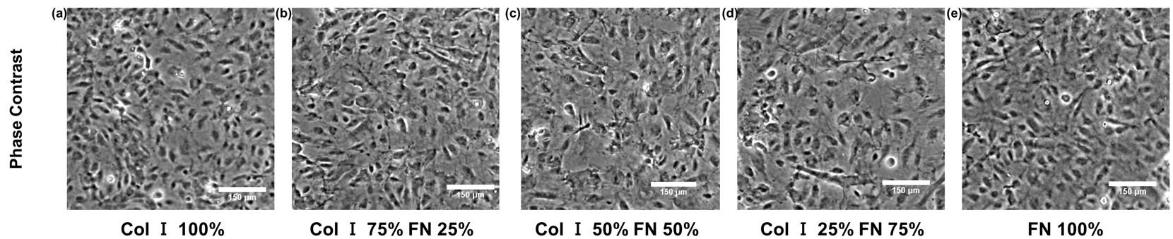
Cropped HUVEC monolayer phase-contrast images (within 651 * 651 Sq. microns) for different Col-I and FN coating concentration ratios: Col-I 100% (A), ColI 75% FN 25% (B), Col-I 50% FN 50% (C), Col-I 25% FN 75% (D), FN 100% (E). Col-I, collagen I; FN, fibronectin; HUVEC, human umbilical vein endothelial cell
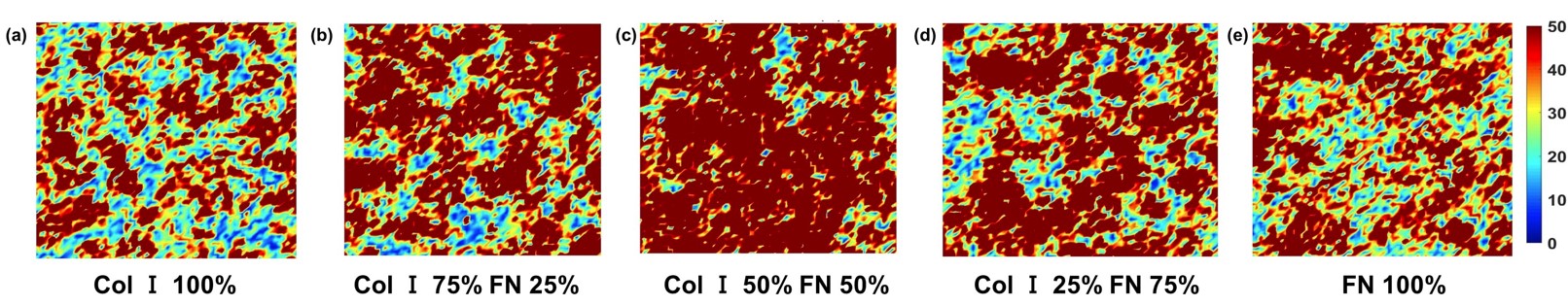
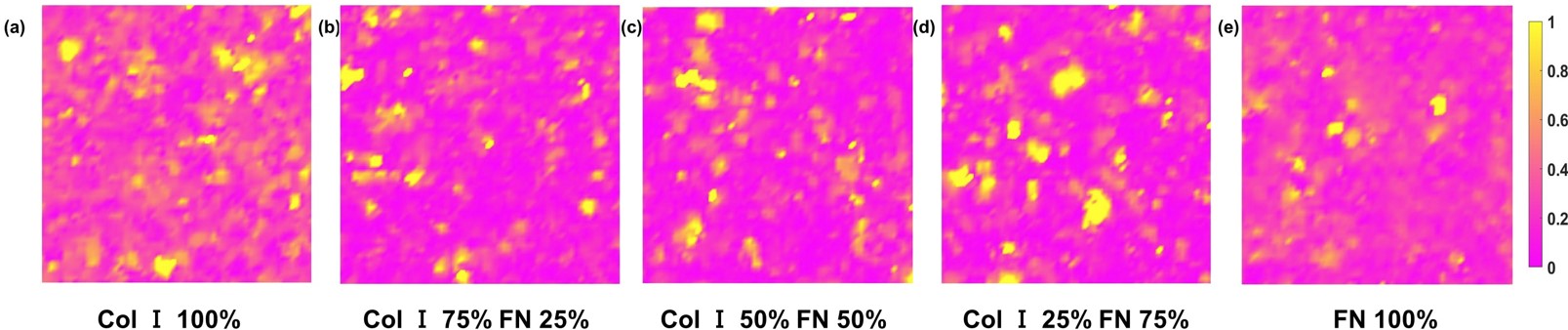
RMS traction (within 651 * 651 Sq. microns cropped section) distributions (Pa) for different Col-I and FN coating concentration ratios: Col-I 100% (A), Col-I 75% FN 25% (B), Col-I 50% FN 50% (C), Col-I 25% FN 75% (D), FN 100% (E)
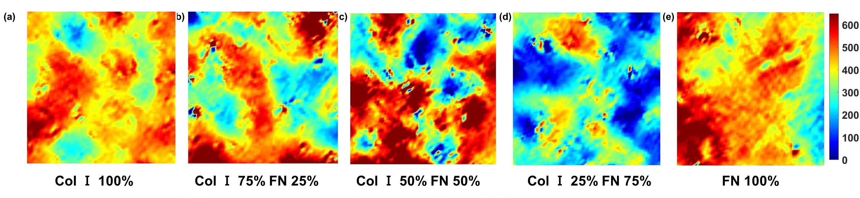
Average normal stress (within 651 * 651 Sq. microns cropped section) distributions (Pa) for different Col-I and FN coating concentration ratios: Col-I 100% (A), Col-I 75% FN 25% (B), Col-I 50% FN 50% (C), Col-I 25% FN 75% (D), FN 100% (E)
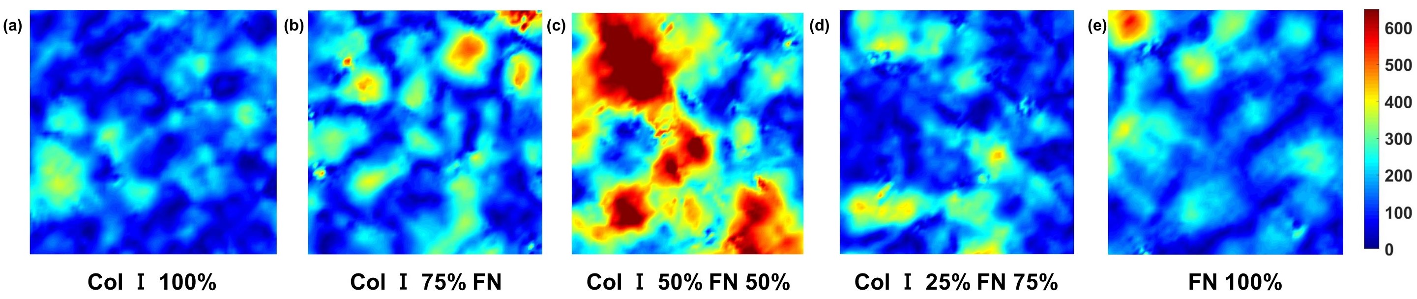
Maximum shear stress (within 651 * 651 Sq. microns cropped section) distributions (Pa) for different Col-I and FN coating concentration ratios: Col-I 100% (A), Col-I 75% FN 25% (B), Col-I 50% FN 50% (C), Col-I 25% FN 75% (D), FN 100% (E)
RMS velocity (within 651 * 651 Sq. microns cropped section) distributions (lm/min) for different Col-I and FN coating concentration ratios: Col-I 100% (A), Col-I 75% FN 25% (B), Col-I 50% FN 50% (C), Col-I 25% FN 75% (D), FN 100% (E)
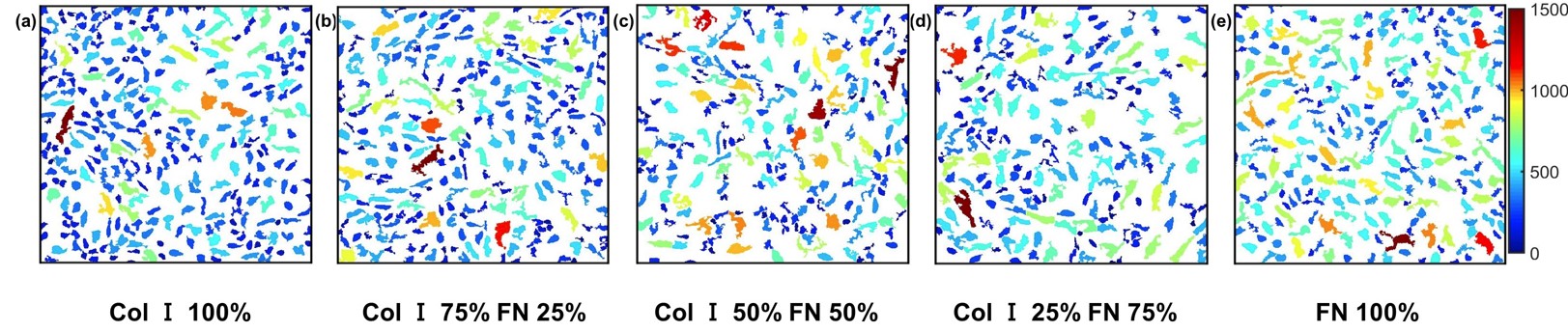
HUVEC area (Sq. microns) (within 651 * 651 Sq. microns cropped section) for different Col-I and FN coating concentration ratios: Col-I 100% (A), Col-I 75% FN 25% (B), Col-I 50% FN 50% (C), Col-I 25% FN 75% (D), FN 100% (E)